Retinoblastoma is an aggressive form of eye cancer that mostly occurs in children from birth to five years old. It is difficult to diagnose because typically, children that young are not seen by an ophthalmologist. Leukocoria, or “white eye” is one of the main symptoms of retinoblastoma. Baylor University and Harvard Medical School researchers believe that leukocoria, or “white eye,” which can be seen in amateur digital photographs, can help get children diagnosed earlier than they would have in the past.
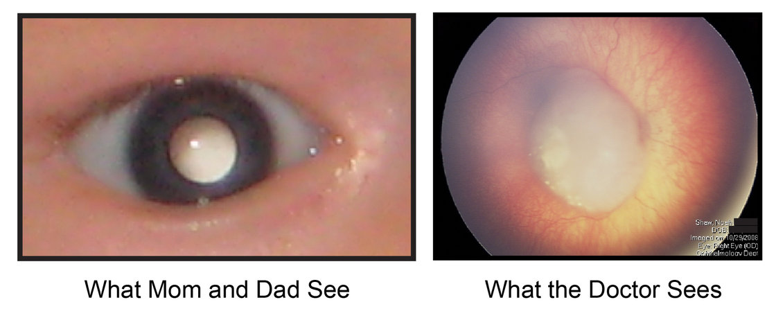
It was known that “white eye” was visible in advanced stages of the cancer, but the research done by Baylor and 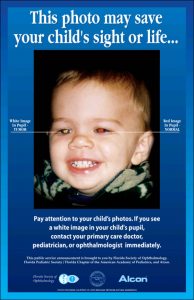 Harvard medical schools has shown that it can be seen much earlier than previously thought. Earlier diagnosis is significant because if retinoblastoma is not found in time it can spread and become fatal. It can also help save the child’s vision. In the United States, children who are treated for retinoblastoma have a 95% chance of being cured. In developing countries, where access to healthcare is limited, the survival rate is much lower. There is increasing access to digital photography in these countries, and it is the hope of researchers that their findings can help save the
  lives of many more children than in the past
Even though digital photography seems to be a good way to detect the cancer, this method is not the standard method currently used by doctors. Researchers are hoping to one day change that, and include digital photography in the screening process.
If you would like to read the original article that inspired this blog post, click here.